Home

Giorno danno sono assetato dii staining protocol Operazione possibile Separare trattenere

DiI and DIO Staining Procedures

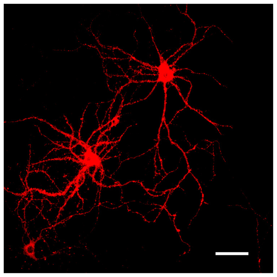
Frontiers | Fluorescent labeling of dendritic spines in cell cultures with  the carbocyanine dye “DiI”
Frontiers | Fluorescent labeling of dendritic spines in cell cultures with the carbocyanine dye “DiI”

Is this DiI staining valid or specific? | ResearchGate

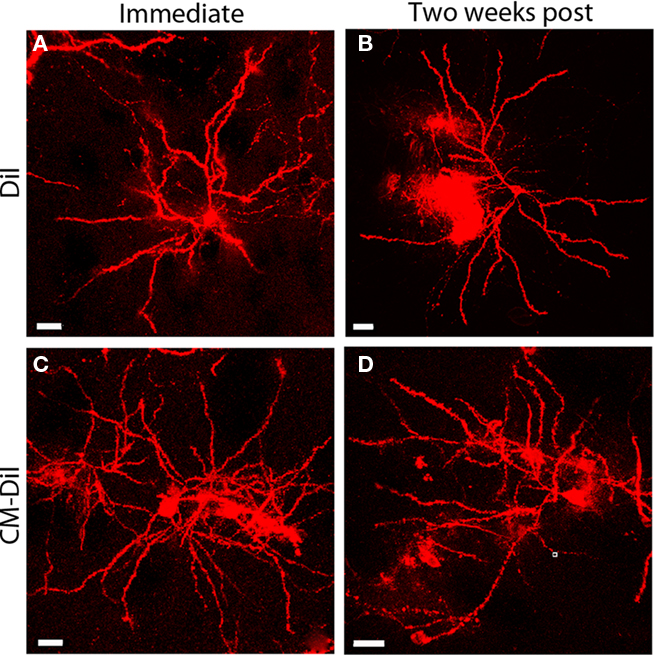
Fluorescent imaging. (A) Vital fluorescent dye staining with CM-Dil did...  | Download Scientific Diagram
Fluorescent imaging. (A) Vital fluorescent dye staining with CM-Dil did... | Download Scientific Diagram

Show your true color: Mammalian cell surface staining for tracking cellular  identity in multiplexing and beyond - ScienceDirect
Show your true color: Mammalian cell surface staining for tracking cellular identity in multiplexing and beyond - ScienceDirect
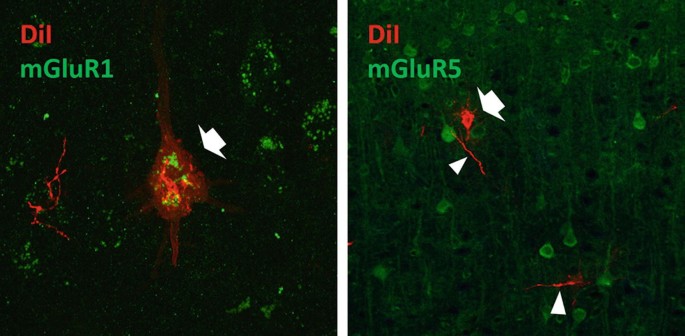
Image Gallery Detail | Thermo Fisher Scientific - US

Co-labeling of Neuronal Cells Using DiI Neuronal Filling and  Immunohistochemistry to Explore Metabotropic Glutamate Receptor Expression  | SpringerLink
Co-labeling of Neuronal Cells Using DiI Neuronal Filling and Immunohistochemistry to Explore Metabotropic Glutamate Receptor Expression | SpringerLink
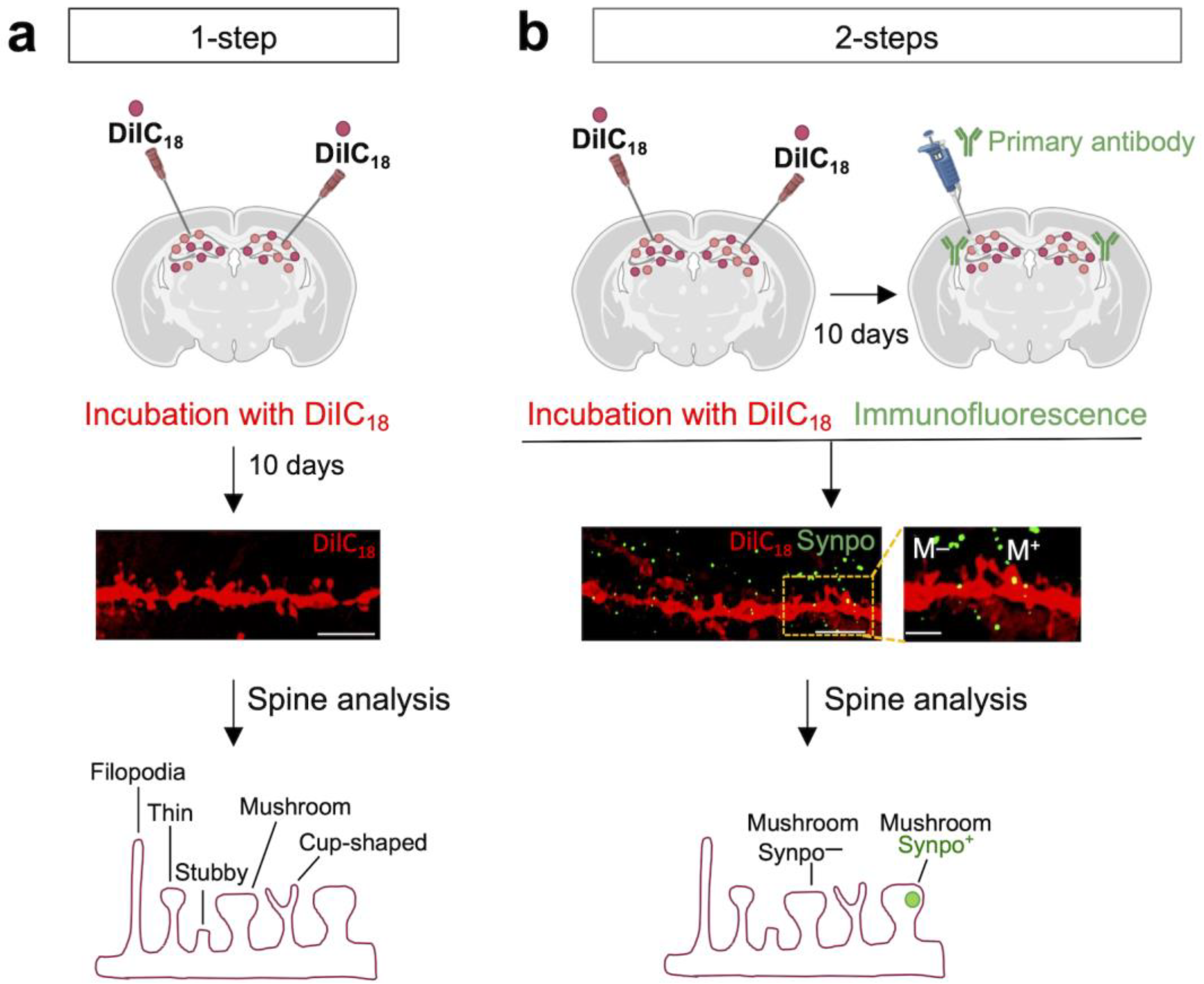
Biomedicines | Free Full-Text | Combined DiI and Antibody Labeling Reveals  Complex Dysgenesis of Hippocampal Dendritic Spines in a Mouse Model of  Fragile X Syndrome
Biomedicines | Free Full-Text | Combined DiI and Antibody Labeling Reveals Complex Dysgenesis of Hippocampal Dendritic Spines in a Mouse Model of Fragile X Syndrome
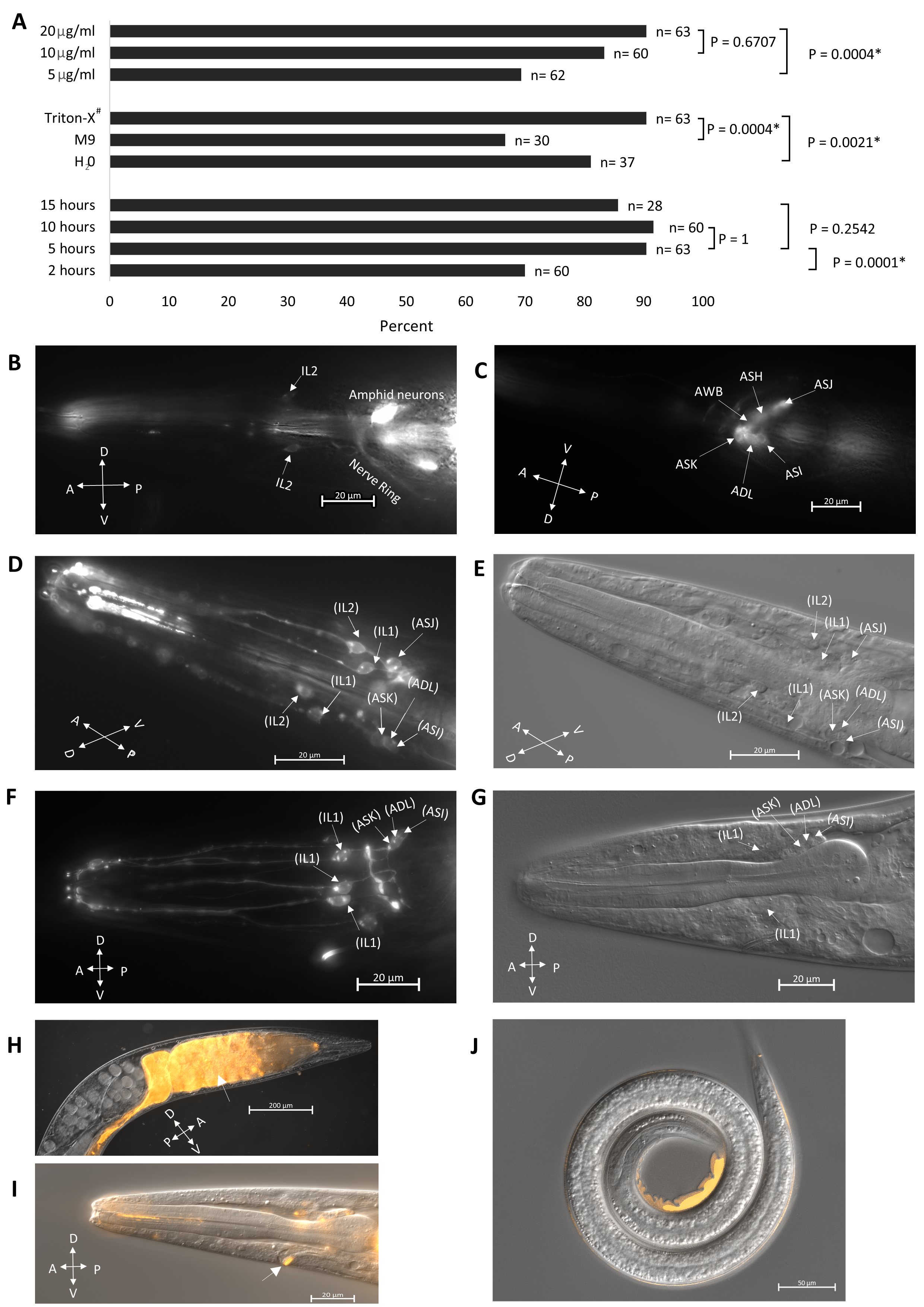
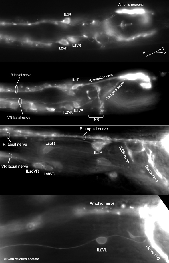
DiI staining of sensory neurons in the entomopathogenic nematode  Steinernema hermaphroditum | microPublication
DiI staining of sensory neurons in the entomopathogenic nematode Steinernema hermaphroditum | microPublication

Is this DiI staining valid or specific? | ResearchGate

Protocol for immunostaining of DiI-labeled cells - 名古屋大学院医学系研究科 細胞生物学分野

A comparative study of PKH67, DiI, and BrdU labeling techniques for tracing  rat mesenchymal stem cells | SpringerLink
A comparative study of PKH67, DiI, and BrdU labeling techniques for tracing rat mesenchymal stem cells | SpringerLink

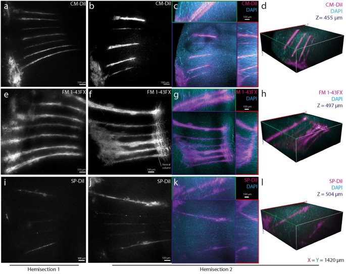
CLARITY-compatible lipophilic dyes for electrode marking and neuronal  tracing | Scientific Reports
CLARITY-compatible lipophilic dyes for electrode marking and neuronal tracing | Scientific Reports

Lipophilic Dye Staining of Cryptococcus neoformans Extracellular Vesicles  and Capsule | Eukaryotic Cell
Lipophilic Dye Staining of Cryptococcus neoformans Extracellular Vesicles and Capsule | Eukaryotic Cell
DiI labeled neurons fixed with varying concentrations of... | Download  Scientific Diagram
DiI labeled neurons fixed with varying concentrations of... | Download Scientific Diagram

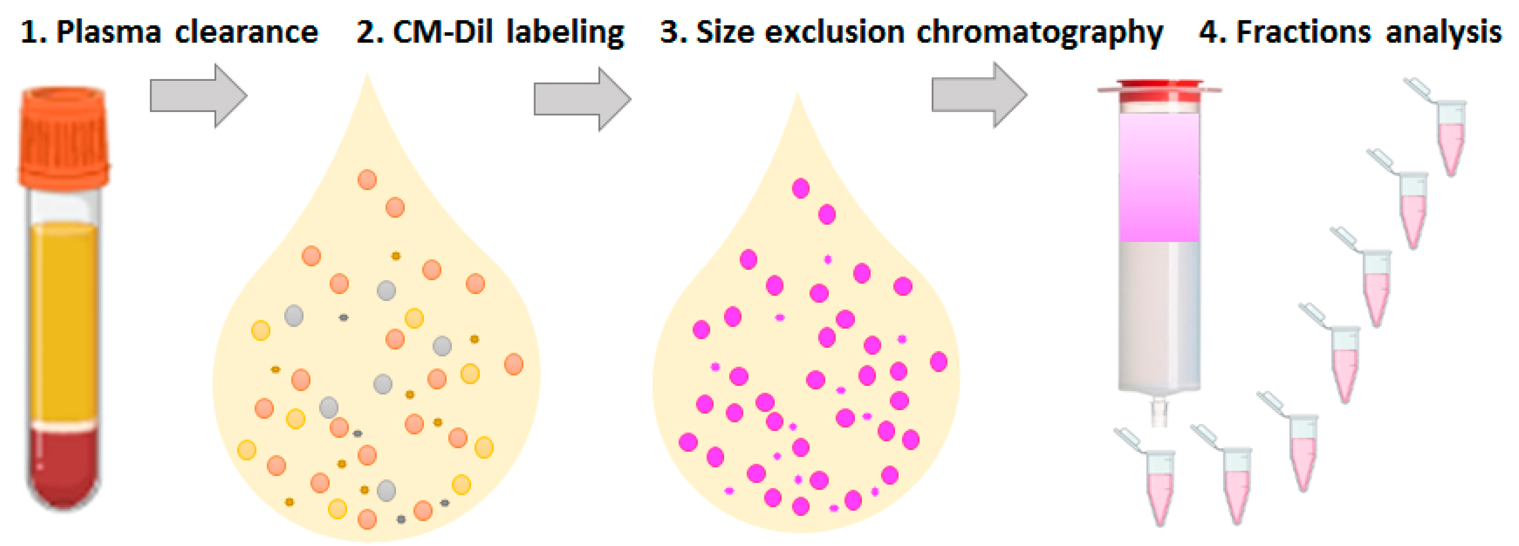
Membranes | Free Full-Text | CM-Dil Staining and SEC of Plasma as an  Approach to Increase Sensitivity of Extracellular Nanovesicles  Quantification by Bead-Assisted Flow Cytometry
Membranes | Free Full-Text | CM-Dil Staining and SEC of Plasma as an Approach to Increase Sensitivity of Extracellular Nanovesicles Quantification by Bead-Assisted Flow Cytometry
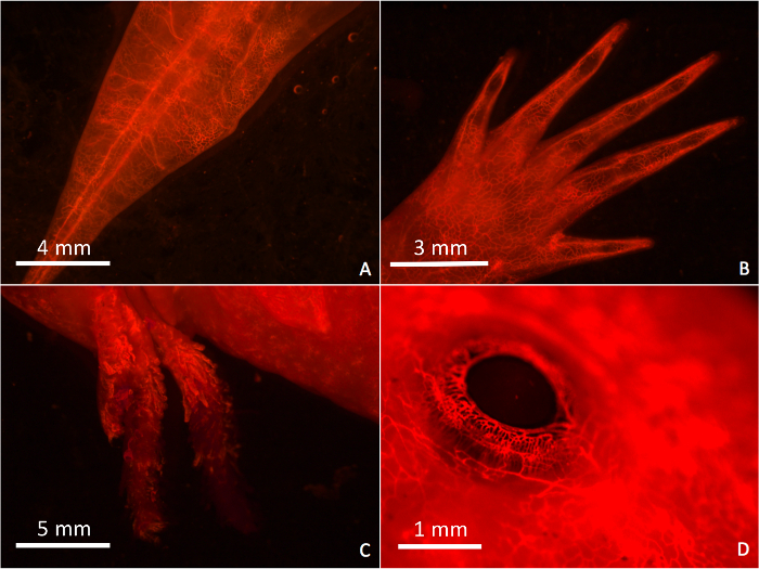
DiI Perfusion as a Method for Vascular Visualization in Ambystoma mexicanum

Frontiers | DiOlistic Labeling of Neurons in Tissue Slices: A Qualitative  and Quantitative Analysis of Methodological Variations
Frontiers | DiOlistic Labeling of Neurons in Tissue Slices: A Qualitative and Quantitative Analysis of Methodological Variations

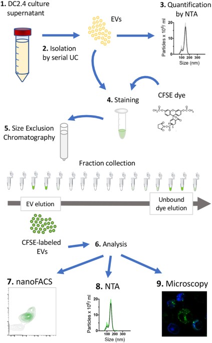
Labeling Extracellular Vesicles for Nanoscale Flow Cytometry | Scientific  Reports
Labeling Extracellular Vesicles for Nanoscale Flow Cytometry | Scientific Reports

Efficient labeling of vesicles with lipophilic fluorescent dyes via the  salt-change method | Exosome RNA
Efficient labeling of vesicles with lipophilic fluorescent dyes via the salt-change method | Exosome RNA

DiI-mediated analysis of pre- and postsynaptic structures in human  postmortem brain tissue | bioRxiv
DiI-mediated analysis of pre- and postsynaptic structures in human postmortem brain tissue | bioRxiv
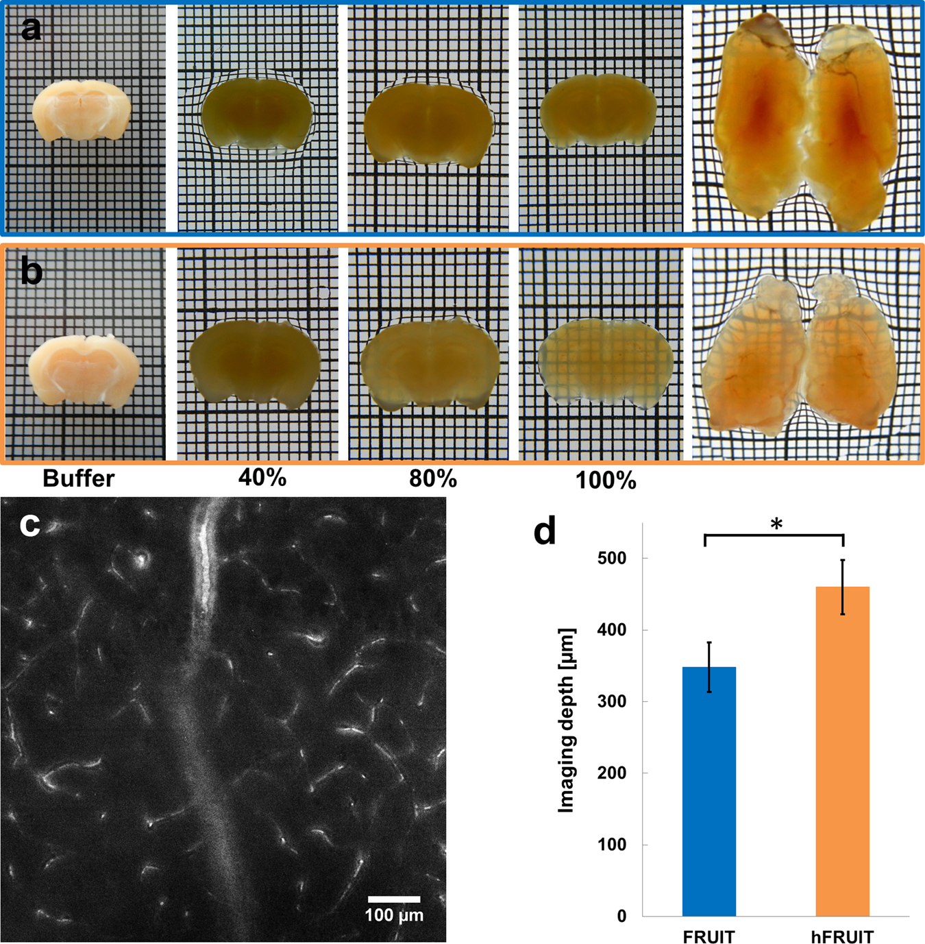
hFRUIT: An optimized agent for optical clearing of DiI-stained adult human  brain tissue | Scientific Reports
hFRUIT: An optimized agent for optical clearing of DiI-stained adult human brain tissue | Scientific Reports